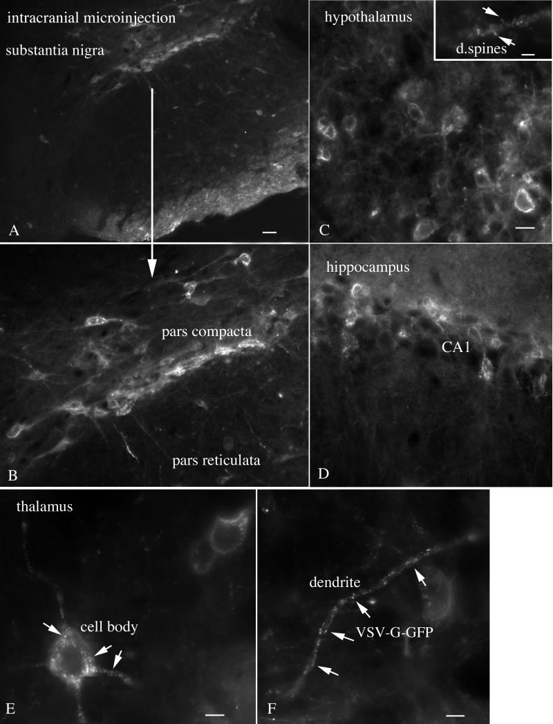
FIG. 4.
Intracranial administration of VSV. Widespread infection, including substantia nigra, hypothalamus, hippocampus, and thalamus, is shown. (A) Two days after intracranial injection of VSV, cells of the pars compacta are infected with VSV. A few cells in the pars reticulata are also infected. Scale bar, 40 μm. (B) Higher magnification of the infected cells of the substantia nigra. Neurons of the medial hypothalamus (C; scale bar, 13 μm), hippocampus (D; scale bar, 20 μm), and thalamus (E and F) are infected with VSV. An inset in panel C shows dendritic spines (arrrows). (E and F) Granule appearance of VSV G-GFP in the cell body and dendrites of thalamic neurons indicative of an early stage of infection. (E) Scale bar, 6 μm. (F) Scale bar, 5 μm.

