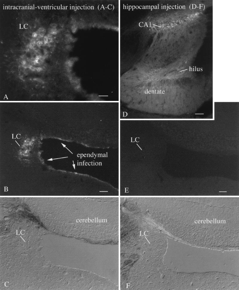
FIG. 5.
Locus coeruleus (LC) selective neurotropism. (A) After intracerebral injections of VSV into rostral brain regions where VSV was found in the ependymal cells of the ventricular system, the LC showed a high level of infection. Scale bar, 40 μm. (B) A lower magnification shows the same field, with infection of the nearby GFP-expressing ependymal cells. Other neurons also adjacent to the ventricular system show only relatively low levels of infection. The LC was infected bilaterally, but only the left LC is shown here. Scale bar, 125 μm. (C) DIC micrograph of the same area shown in panel B. (D) A small injection into the hippocampus shows infection of CA1 and the dentate gyrus and hilus. On other sections, not shown here, part of CA3 was also infected. Scale bar, 225 μm. (E) In the absence of VSV in the ventricular system, no infection of the LC is detected. Scale bar, 125 μm. (F) DIC micrograph of the same field shown in panel E.

