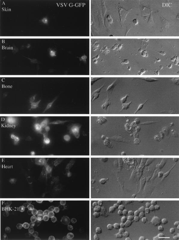
FIG. 8.
Multiple cell types are infected by VSV. Primary cultures were made from skin (A), brain (B), bone (C), kidney (D), and heart (E). (F) BHK-21 cells that are used to propagate the virus were used for the purpose of comparison. Cultures were fixed with paraformaldehyde 8 h p.i. The expression of VSV G-GFP is shown on the left, and the same microscope field is shown on the right side with DIC. Scale bar, 30 μm.

