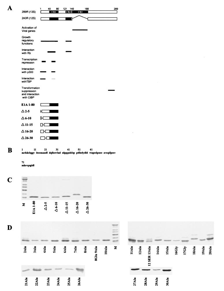
FIG. 1.
(A) Schematic of E1A 289R and 243R proteins, their domain structures, and associated biological activities. Below are schematics of E1A 1-80 and E1A 1-80 deletion polypeptides. (B) Sequence of the first 80 amino acids of E1A (E1A 1-80). (C) Purity of E1A 1-80 deletion mutants. One-microgram amounts of polypeptide were resolved on 15% polyacrylamide gels, stained with SYPRO orange (Molecular Probes), and visualized by blue-green fluorescence on a STORM 840 PhosphorImager. The lowest band of the marker lane (M) is 10 kDa. (D) Purity of E1A 1-80 amino acid substitution mutants. One-microgram amounts of the E1A 1-80 mutants were analyzed as described for panel C above.

