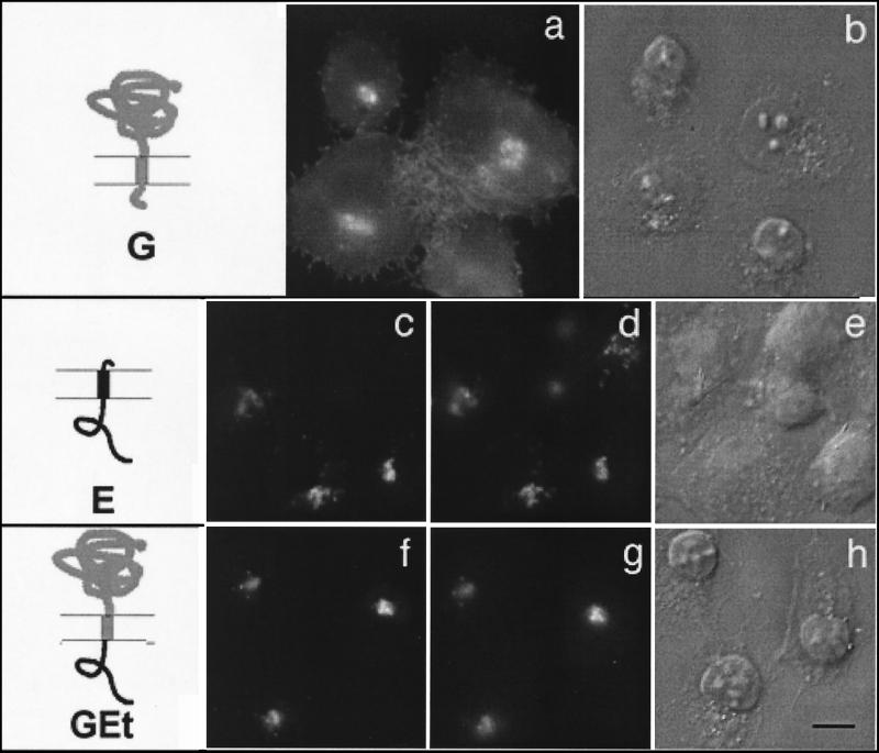
FIG. 3.
Cytoplasmic tail of IBV E is sufficient to retain a reporter protein in the Golgi complex. BHK cells expressing wild-type VSV G (a and b), wild-type IBV E (c to e), or a chimeric protein consisting of the lumenal head and transmembrane domain of VSV G and the cytoplasmic tail of E (GEt; f to h) were fixed for immunofluorescence, permeabilized, and stained with anti-G (a) or double labeled with anti-E (c and f) and anti-GM130 (d and g) antibodies. Secondary antibodies were fluorescein-conjugated donkey anti-rabbit IgG and Texas Red-conjugated goat anti-mouse IgG. DIC images of the labeled cells are shown in panels b, e, and h. The diagrams indicate IBV E sequence in black and VSV sequence in gray. Bar, 10 μm.

