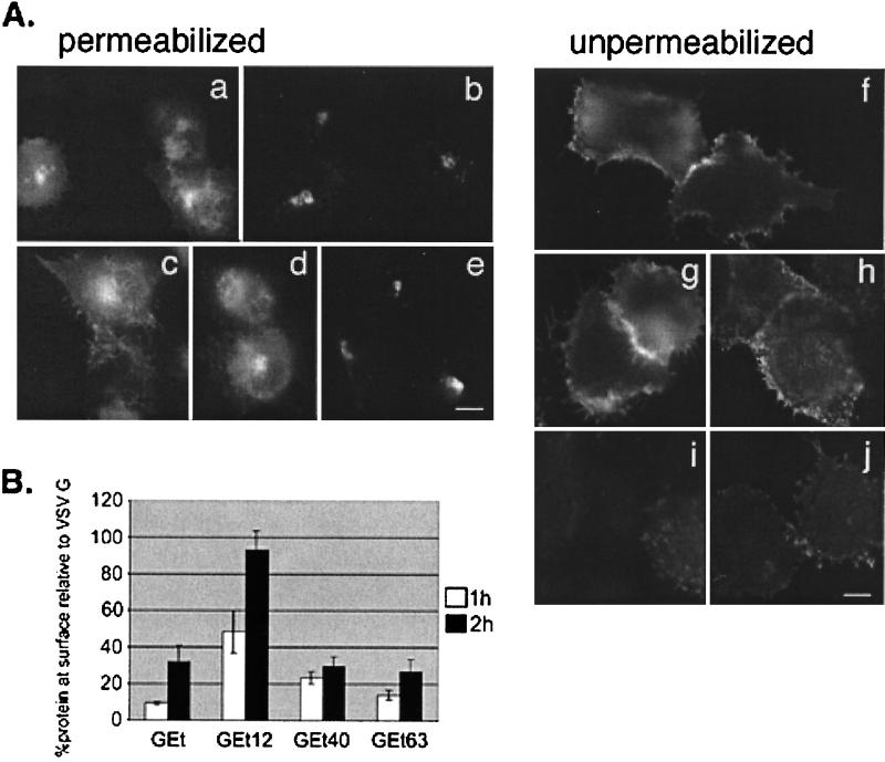
FIG. 6.
Golgi targeting information in the IBV E protein is between residues 12 and 63 of the cytoplasmic tail. (A) BHK cells expressing VSV G (a and f), GEt (b and i), GEt12 (c and g), GEt40 (d and h), or GEt63 (e and j) were fixed for immunofluorescence and permeabilized (a to e) or fixed and left unpermeabilized (f to j) and stained with an antibody to the ectodomain of VSV G. Secondary antibody was Texas Red-conjugated goat anti-mouse IgG. (B) BHK cells expressing GEt, GEt12, GEt40, or GEt63 were pulse-labeled with [35S]methionine-cysteine and chased for 1 h (white bars) or 2 h (black bars). Intact cells were incubated with polyclonal anti-VSV antibody for 2 h at 4°C. After cell lysis, antibody complexes were collected, and supernatants were incubated with anti-VSV to immunoprecipitate intracellular proteins. Samples were run on SDS-PAGE, and quantitation of surface and internal proteins was performed. A ratio of the amount of surface to internal protein was calculated for GEt and truncation derivatives. Shown on the graph is this ratio taken as a percentage of the surface/internal ratio of wild-type VSV G protein at the corresponding time point. Each value represents an average of three experiments ± SEM.

