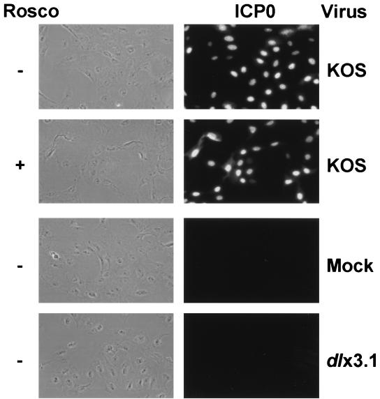
FIG. 4.
Rosco does not affect the nuclear localization of ICP0. Vero cells were pretreated with CHX (50 μg/ml) for 1 h, mock infected or infected with 5 PFU per cell of KOS or dlx3.1 (an ICP0 null mutant), and incubated for 6 h in the presence of CHX. Viruses used are indicated on the right of the figure. Cells were then released from the CHX block in the absence (−) or presence (+) of Rosco for 6 h, as indicated. At the end of the second 6-h treatment period, cells were washed, fixed, permeabilized, and incubated with a primary mouse monoclonal antibody against ICP0 (1). Primary antibody was detected with a fluorescein isothiocyanate-conjugated rabbit anti-mouse antibody. Monolayers were viewed by phase-contrast (left column) and fluorescence microscopy (right column) at ×400 magnification, and cells were photographed with a digital camera.

