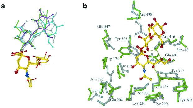
FIG. 4.
Sialyllactose modelling. (a) Four sialyllactose ligands taken from protein-ligand complexes have been superimposed on their sialic acid moieties, shown in atom coloring: yellow carbons, blue nitrogens, and red oxygens. The galactose and glucose residues are colored grey for the sugars from their complex with the influenza virus hemagglutinin, cyan for leukoagglutinin, magenta for sialoadhesin, and green for murine polyomavirus. (b) Sialyllactose modelled into the binding site of NDV HN. The side chains of the unliganded crystallographic structure are shown in grey, and the modelled structure is shown in green.

