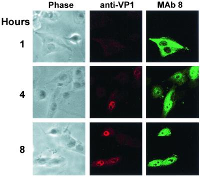
FIG. 3.
Exposure of the VP1 unique region on purified capsids injected into cells. Full virus capsids were injected into cells, which were then incubated for 1, 4, or 8 h at 37°C before fixation. The cells were incubated with rabbit anti-VP1 unique region followed by TxR-conjugated anti-rabbit IgG. Capsids were detected using MAb 8 and FITC-conjugated anti-mouse IgG. Confocal images show a 1-μm section from the center of each cell, while phase-contrast images of the same cells are also shown.

