
FIG. 5.
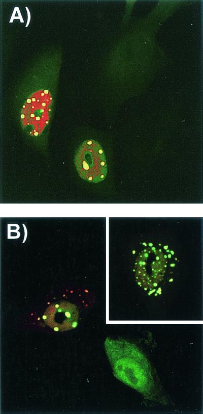
Detection of CPV replication and of capsid protein localization in cells injected simultaneously with anti-VP1-2-13 and the CPV infectious plasmid and then incubated for 24 h at 37°C. (A) Expression of NS1 in cells coinjected with anti-VP1-2-13 and a CPV infectious plasmid. The anti-VP1-2-13 was detected with an FITC-labeled anti-mouse IgG (green), and NS1 was detected with a TxR-labeled anti-NS1 IgG (red). (B) Localization of CPV capsid proteins in cells coinjected with anti-VP1-2-13 and a CPV infectious plasmid. The injected MAb was detected with FITC-labeled anti-mouse IgG (green), and capsid proteins were detected with a specific rabbit anticapsid IgG followed by TxR-labeled anti-rabbit IgG (red). The inset shows another cell subjected to the same treatment.