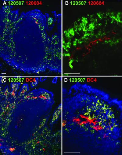
FIG. 7.
Comparison of DC-SIGN and DC-SIGNR expression in human Peyer's patches. Human Peyer's patches were doubly labeled with mouse MAbs specific for DC-SIGN (120507; green) or DC-SIGNR (120604; red) (A and B). The staining by MAb DC4 (recognizing both DC-SIGN and DC-SIGNR; red) and DC-SIGN-specific staining by MAb 120507 (green) were compared (C and D). The nucleus was stained with DAPI (blue). To examine the presence of DC-SIGN+/DC-SIGNR+ cells found in the endothelium of the dome region in A, confocal microscopy analysis was carried out using a 40× objective lens (B). Images were captured using 10× (A and C) or 40× (B and D) objective lenses. Close-up of subepithelial dome region in panel C is shown in panel D.

