FIG. 4.
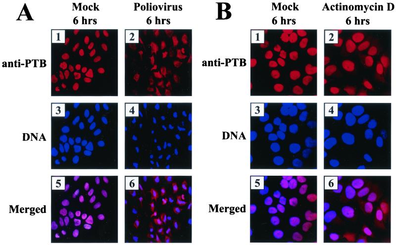
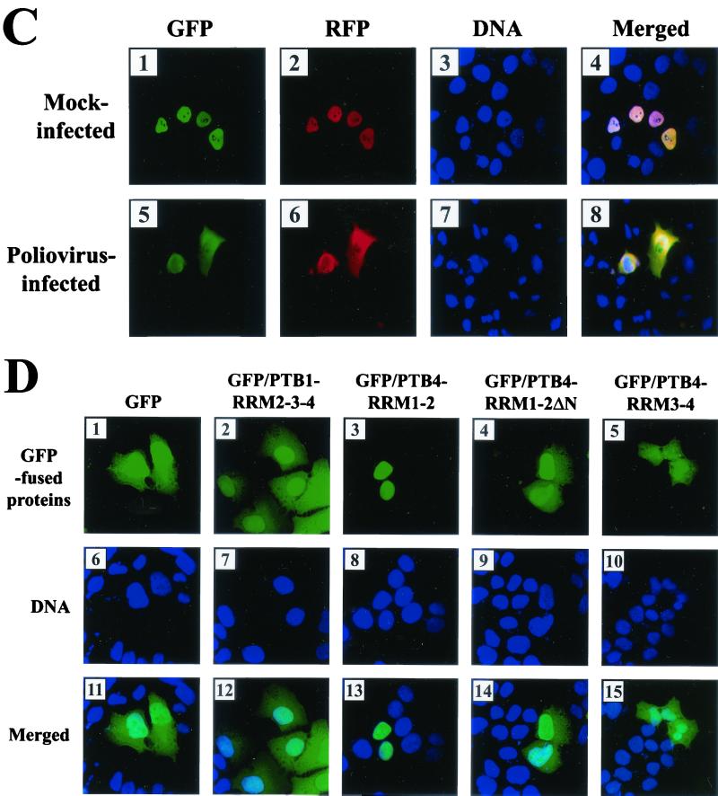
Subcellular localization of PTB and its derivatives. (A) Effect of poliovirus infection on PTB localization. HeLa cells were mock infected or infected with poliovirus for 6 h and then stained with antibody directed against PTB. The top panels show cells examined with a TRITC filter, the middle panels show Hoechst 33258 staining of the same field with a 4",6"-diamidino-2-phenylindole (DAPI) filter, and the bottom panels show the merged image of the TRITC and Hoechst images. (B) Effect of transcription blockage by actinomycin D on PTB localization. Uninfected HeLa cells were incubated without or with actinomycin D (5 μg/ml) for 6 h and then fixed and stained with anti-PTB monoclonal antibody. (C) HeLa cells were transfected with plasmids expressing GFP/PTB4/RFP and then mock infected or infected with poliovirus for 6 h from 40 h posttransfection. GFP fluorescence and RFP fluorescence were visualized with a fluorescein isothiocyanate (FITC) filter and a TRITC filter, respectively. DNA was examined with a DAPI filter after Hoechst 33258 staining. Panels 4 and 8 are merged images of FITC, TRITC, and Hoechst images. (D) Subcellular localization of PTB fragments. HeLa cells were transiently transfected with plasmids expressing GFP (panels 1, 6, and 11), GFP fused with PTB1-RRM2-3-4 (panels 2, 7, and 12), PTB4-RRM1-2 (panels 3, 8, and 13), PTB4-RRM1-2 lacking NLS (panels 4, 9, and 14), and PTB4-RRM3-4 (panels 5, 10, 15). GFP fluorescence was visualized with an FITC filter. DNA was stained with Hoechst 33258 and examined with a DAPI filter.


