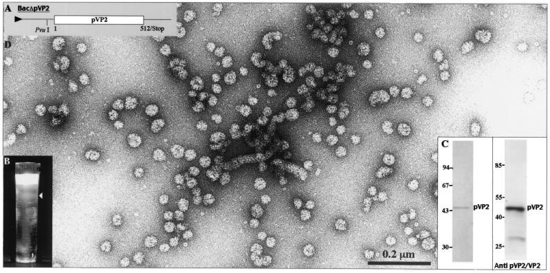
FIG. 3.
Analysis of the structures produced by BacΔpVP2. (A) Map of the construct encoding pVP2. Sf9 cells were infected with BacΔpVP2 and treated with Freon 113 as described in Materials and Methods. (B) After an 18-h centrifugation in CsCl at 100,000 × g, the gradient was illuminated with white light and photographed. (C) The left panel shows SDS-PAGE analysis and Coomassie blue staining of the material isolated from the fuzzy band present in the CsCl gradient. The right panel shows a Western blot analysis with an anti-pVP2/VP2 antibody. (D) Material collected from the band present in the gradient was negatively stained with 1% uranyl acetate. Large numbers of small capsids and some flexible tubes were vizualized.

