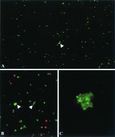
FIG. 6.
Visualization of individual fluorescent VLPs (A and B) A1-μl volume of purified VLPs was directly examined under a optical microscope (A) or mixed with red-fluorescent latex beads and examined under a confocal microscope (B). (C) VLPs were examined, after addition of an anti-VP2 antibody under a confocal microscope.

