FIG. 7.
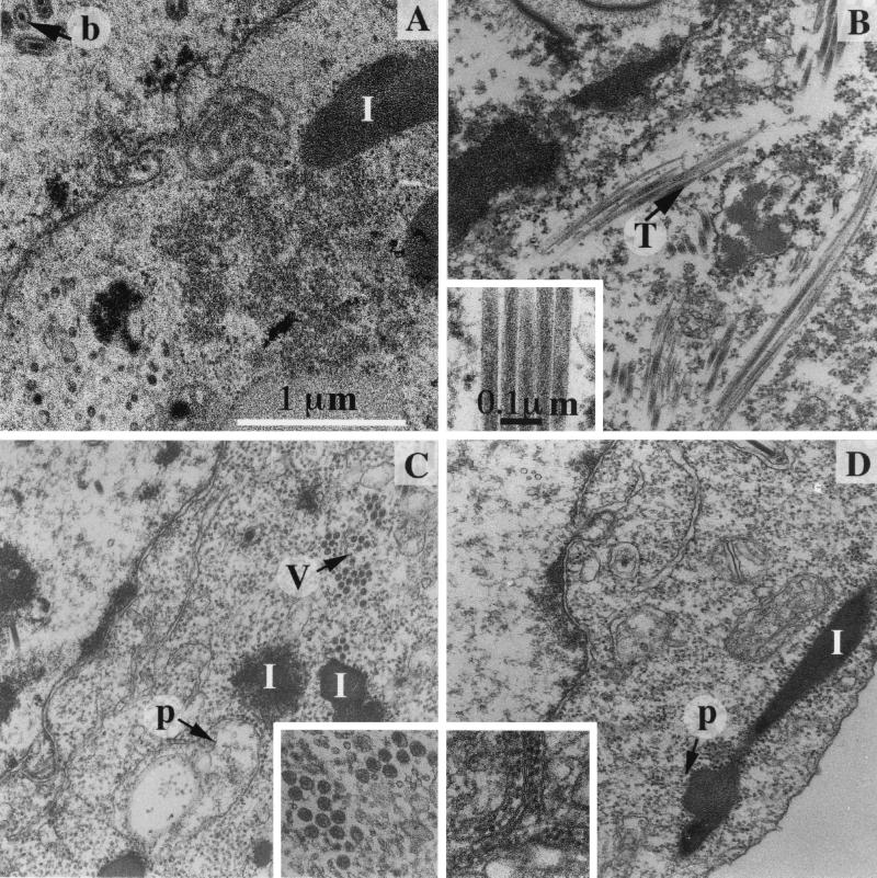
Micrographs of thin sections of Sf9 cells infected by different recombinant baculoviruses. (A) The polyhedrin-negative baculovirus. Baculoviruses are assembled in the nucleus (b). In the cytoplasm, the main consequence of the baculovirus infection is the accumulation of inclusions with a more or less regular shape (I). (B) BacΔIBDA; (C) BacΔIBDA-GFP; (D) BacΔpVP2. The characteristic structures formed by the polyprotein-derived constructs are inserted in the corresponding micrographs: 50-nm-diameter tubules (T), VLPs (V), and isometric particles often associated with membranes (p). Tubules are occasionally observed in cells infected with BacΔIBDA-GFP (data not shown). Small numbers of isometric particles are also identified in cells infected by BacΔIBDA and BacΔIBDA-GFP, suggesting that pVP2 may self-assemble in all cases. Magnifications are identical in all micrographs and inserts.

