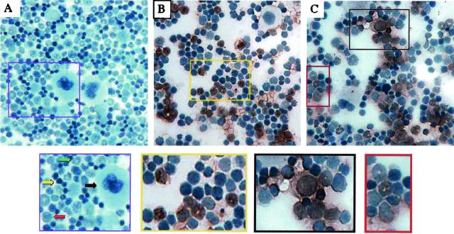
FIG. 4.
Immunohistochemical staining for SL3 SU protein in bone marrow cells from uninfected NIH/Swiss mice (A) or from mice infected with SL3 (B) or SL3ΔMyb5 (C). Bone marrow was collected from the femur at 3 weeks p.i., deposited onto glass slides by cytocentrifugation, and stained immunohistochemically for SL3 SU (reddish-brown precipitate). Cells were then counterstained with hematoxylin. The enlarged image from panel A (purple box) shows four morphologically distinct types of cells in uninfected marrow: (i) very large cells with abundant cytoplasm and irregular nuclei, characteristic of megakaryocytes (black arrow); (ii) medium-sized cells with large, often eccentric nuclei and relatively abundant cytoplasm, a morphology consistent with immature myeloid precursors (red arrow); (iii) medium-sized cells with crescent- or ring-shaped nuclei typical of maturing cells of the granulocyte lineage (green arrow); and (iv) small cells with scant cytoplasm and condensed nuclei, characteristic of a heterogeneous population including myeloid precursors, erythroid precursors, and mature and maturing lymphocytes (yellow arrow). The enlarged image from panel B (yellow box) shows SL3 infection predominantly in immature and maturing granulocytes with ring-shaped nuclei. The enlarged images from panel C show SL3ΔMyb5 infection predominantly in immature myeloid precursors (black box) and maturing granulocytes (red box).

