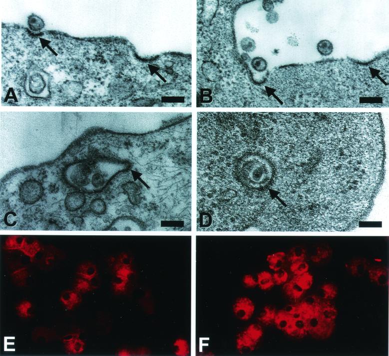
FIG. 6.
Virus particles are taken up via clathrin-coated pits by immature and mature DCs. Human immature (A, B, and D) and mature (C) DCs were first incubated for 15 min at 4°C with AT-2 SIV (30 ng of SIV p27 Ag/105 cells). After a further 1 min (C), 5 min (A), or 120 min (B and D) at 37°C, the cells were washed, fixed, and processed for electron microscopy. Whole, structurally intact virus can be spotted bound to coated pits (A and B, arrows) and localized in coated vesicles close beneath the plasma membrane surface (C and D, arrows). Magnification and scale bars: ×50,000/200 nm (A); ×41,000/240 nm (B); ×47,000/210 nm (C); and ×80,000/120 nm (D). Cytospin-adhered human immature (E) and mature (F) DCs were fluorescently labeled for clathrin (X-22) and revealed comparable amounts of clathrin-coated structures.

