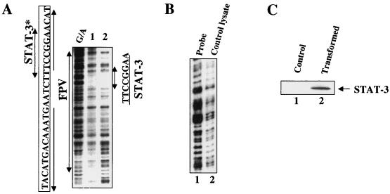
FIG. 4.
DNase I protection analysis of STAT-3 binding. (A) Samples were analyzed in the presence of the enhancer 1 probe (25,000 cpm; labeled at nt 1308). Lanes 1 and 2 contained 50 and 100 μg of STAT-3 protein, respectively. G/A, sequencing ladder. The STAT-3 binding site is shown (nt 1111 to 1121) with a double-headed arrow indicating an 8-bp palindrome. ∗, previously designated as the PBF binding site (28). (B) DNase I protection assay. Lane 1, probe; lane 2, probe incubated with bacterial lysates lacking STAT-3 expression vector. (C) Western blot analysis. Bacterial lysates lacking STAT-3 vector (lane 1), doubly transformed with STAT-3 and JAK vectors (lane 2) were subjected to SDS-7.5% PAGE followed by immunoblot analysis using anti-STAT-3 polyclonal antibodies.

