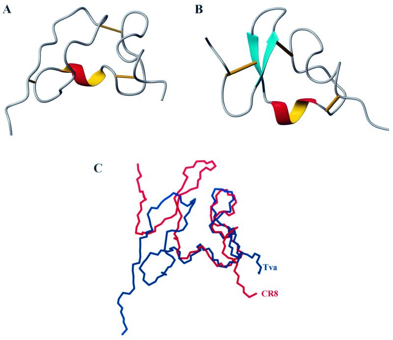
FIG. 2.
Structure comparison of the Tva LDL-A module and LRP CR8. Left, N terminus; right, C terminus. Cyan, β-sheet; red and yellow, α-helix. Bottom right, calcium-binding site; straight lines, three disulfide bonds. (A) Ribbon representation of the final structure of the Tva LDL-A module, based on the 20 best structures shown in Fig. 1. (B) Ribbon representation of the three-dimensional solution structure of CR8. (C) Superimposition of the backbone of the Tva LDL-A module (blue) and that of CR8 (red).

