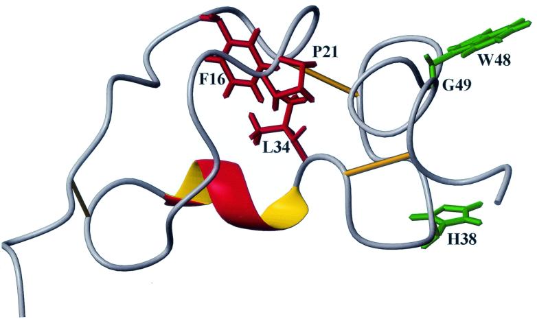
FIG. 6.
Molecular model of Tva as the RSV-A receptor. The side chains of the putative ligand contact residues (green) are surface exposed, whereas the side chains of the residues involved in the formation of the hydrophobic core (red) are buried. In this model, it is proposed that the conformation of the more flexible N terminus of Tva is rigidified upon EnvA binding (induced fit).

