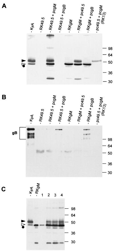
FIG. 2.
Western blot analyses of RK13, RK49.5, and RKgM cells transfected with pc49.5, pcgM, or pcgB. Forty-eight hours after transfection, cells were harvested and lysed, and protein extracts were separated by SDS-10% PAGE followed by Western blotting using anti-gM antibody P18/A8 (A and C) or anti-gB antibody 3F8 (B). Cells lines used for the transfections are given. The gM precursor (circle), the high-mannose gM (open arrowhead), and the fully glycosylated gM (solid arrowhead) are indicated. Individual cell clones which constitutively expressed the UL49.5 protein were identified by the presence of processed gM after transfection of pcgM (C). Cell line 4 was chosen for further studies and termed RK49.5. Mature and immature gM are indicated by solid and open arrowheads, respectively (A and C); gB-specific bands are marked by a bracket (B). The sizes of a prestained molecular weight marker (SeaBlue; Novex) are given in thousands.

