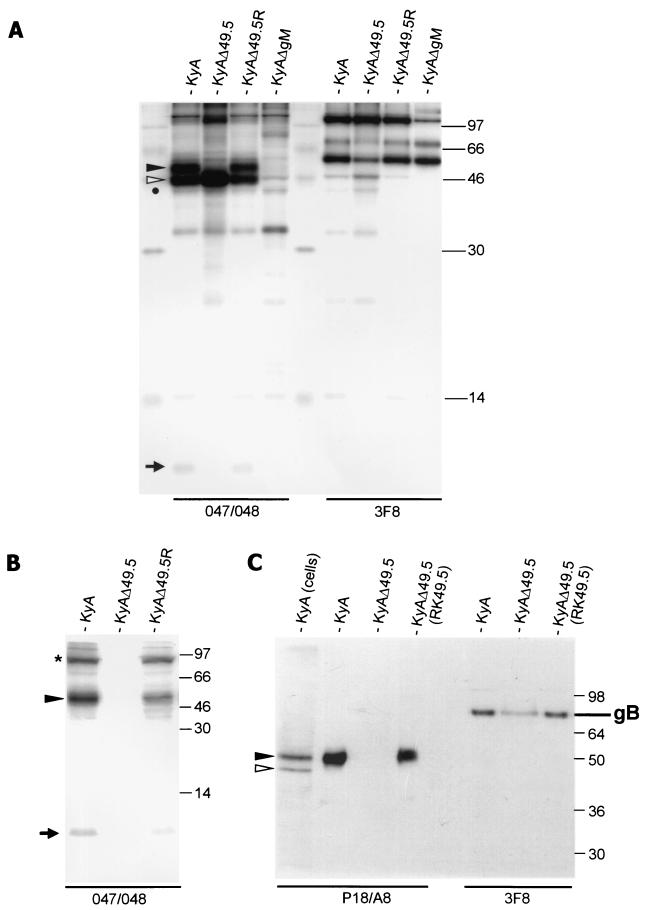
FIG. 3.
(A) RIPA analysis of RK13 cells infected with wild-type KyA, the UL49.5- (gene 10)-negative KyAΔ49.5 mutant, the UL49.5 revertant virus, or gM-negative KyAΔgM (27). (B) RIPA of purified radiolabeled KyA, KyAΔ49.5, or KyAΔ49.5R virions The antibodies used for precipitations were anti-gB antibody 3F8 and the anti-gM polyclonal antibodies 047 and 048. Mature and immature gM are indicated by full and open arrowheads, respectively; 2-mercaptoethanol-resistant gM dimers (24, 27) are indicated by an asterisk. The putative UL49.5 (gene 10) product is indicated by an arrow. The sizes of a molecular weight marker (14C marker; Gibco-BRL) are given in thousands. (C) Western blot analysis of purified KyA and KyAΔ49.5 virions prepared from RK13 or RK49.5 cells by using anti-gM MAb P18/A8 or anti-gB MAb 3F8. Whereas the 72,000- to 75,000-Mr large subunit of gB could be detected with MAb 3F8 in all virion preparations, gM was detected in KyA virions and KyAΔ49.5 virions prepared from RK49.5 cells. gM was absent from UL49.5-negative virions prepared on RK13 cells. Samples were heated for 2 min at 56°C for detection of gM and for 3 min at 95°C for detection of gB. In the first lane, KyA-infected RK13 cells were loaded. The sizes of a prestained molecular weight marker (SeaBlue; Novex) are given in thousands.

