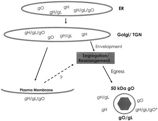
FIG. 8.
Model of gH-gL-gO complex assembly and trafficking in CMV-infected cells. The appearance of various gH, gL, and gO complexes is illustrated temporally from top to bottom. Proteins denoted with a slash (e.g., gH/gL) are held together by disulfide linkages. The glycoproteins are cotranslationally inserted into the ER. Subsequently, glycosylated proteins travel to the Golgi apparatus and then to the TGN. gH, gL, and gO accumulate in Golgi apparatus-derived vesicles, although some gH-gL-gO appears on the surface of infected cells. Cell surface tripartite complexes may or may not recycle via endocytosis (?). Multiple TGN-derived complexes likely incorporate into virions during envelopment. Rearrangement of gH-gL-gO complexes results in the emergence of previously undetected forms of gO, including gO-gL heterodimers. Subsequent events in viral egress are not well characterized. Bold letters indicate gO complexes that arise between the Golgi/TGN and the point of virion collection. gH-gL-gO∗, tripartite complex in which gO contains O-glycans.

