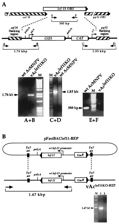
FIG.2.
Confirmation of BACmid constructs vAclef11KO and vAclef11KO-REP by PCR analysis. (A) The strategy for PCR analysis of the lef-11 locus in BACmid vAclef11KO is indicated by the positions of primer pairs (arrows and brackets). The top diagram shows the structure of the wild-type (wt) lef-11 locus, and the lower diagram shows the structure of the BACmid vAclef11KO. To confirm the insertion of the polyA-GUS-CAT-ie1 cassette in BACmid vAclef11KO, primer pairs A+B and C+D were used to examine the recombination junctions by PCR analysis. For each primer pair, one primer corresponded to sequences within the inserted sequence (the GUS or CAT ORF), and the second primer was from the baculovirus genome, just outside the homologous flanking sequences used for recombination. Primer pair E+F was designed to amplify a fragment from within the lef-11 ORF and was used to confirm the absence of the lef-11 ORF in vAclef11KO. The sizes of expected PCR amplification products are shown below each primer pair on the diagram, and the panels below show the agarose gel electrophoresis results of each PCR, with the sizes of PCR products indicated beside an arrowhead. Primer pairs used for each PCR analysis are indicated below each panel and template DNAs are indicated above the panels. M, DNA size markers. (B) Analysis of the polyhedrin locus in BACmid vAclef11KO-REP. PCR analysis was used to confirm the insertion of a cassette containing the lef-11 ORF under the control of the wild-type lef-11 promoter from plasmid pFastBAClef11-REP, into BACmid vAclef11KO. The relative location of the primer pair used to confirm the insertion of the lef-11 gene is shown below the diagram of the resulting BACmid (vAclef11KO-REP). The panel below shows an ethidium bromide-stained agarose gel with the expected 1.47-kbp DNA product of PCR amplification from vAclef11KO-REP (lane 1). A similar PCR amplification from a negative control (wild-type AcMNPV) is also shown (lane 2).

