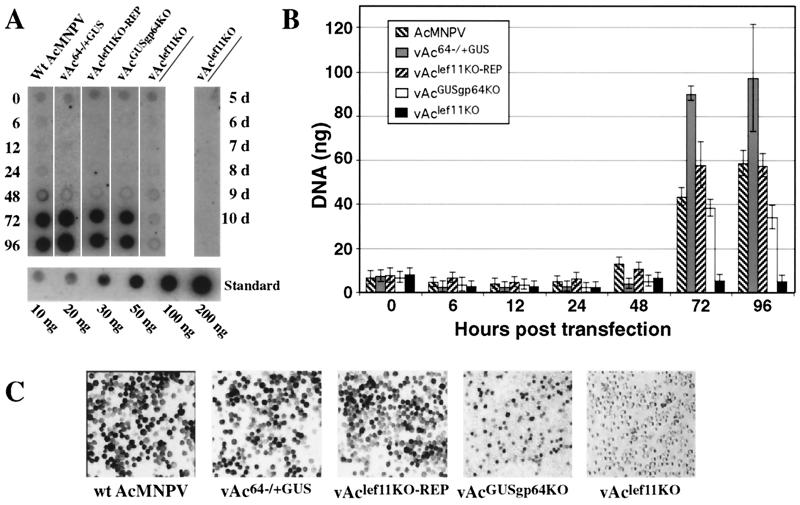
FIG. 5.
Analysis of AcMNPV DNA replication in Sf9 cells transfected with lef-11-null and control BACmids. (A) Sf9 cells were transfected with either wild-type (Wt) AcMNPV, control BACmid (vAc64−/+GUS), lef-11 repair BACmid (vAclef11KO-REP), gp64-null control BACmid (vAcGUSgp64KO), or lef-11-null BACmid (vAclef11KO) DNA (lanes 1 to 5, respectively), and total cellular DNAs were isolated at various times (0 to 96 h) posttransfection. Viral DNA replication was detected by Southern dot blot hybridization with total AcMNPV DNA as a 32P-labeled hybridization probe. Cells transfected with the lef-11-null BACmid (vAclef11KO) were also examined after an extended period (right lane, 5 to 10 days). A standard curve of AcMNPV DNA is shown below (10 to 200 ng of DNA). (B) Quantitative analysis of BACmid DNA replication by Southern dot blot analysis. Three replicates of each virus and time point were examined as shown in panel A, and DNA was measured by PhosphorImager analysis. Bars represent the average of three dot blot samples, and error bars represent standard deviation. (C) Sf9 cells transfected with each of the indicated DNAs (wild-type AcMNPV, vAc64−/+GUS, vAclef11KO-REP, vAcGUSgp64KO, or vAclef11KO) were stained for GUS activity at 4 days posttransfection.

