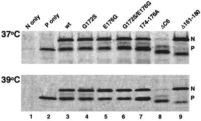
FIG. 2.
Immunoprecipitation analysis of N-P interaction in cells transiently expressing N and P. MVA-T7-infected HEp-2 cells were transfected with pN and different pP protein expression plasmids under the control of T7 promoters and incubated for 16 h at 37°C (upper panel) or 39°C (lower panel). The proteins were radiolabeled with [35S]Cys and [35S]Met (100 μCi/ml) in DMEM deficient in cysteine and methionine for 4 h, immunoprecipitated by anti-P monoclonal antibodies, separated on a 15% polyacrylamide gel, and exposed to Kodak BioMAX film. The positions of N and P are indicated on the right.

