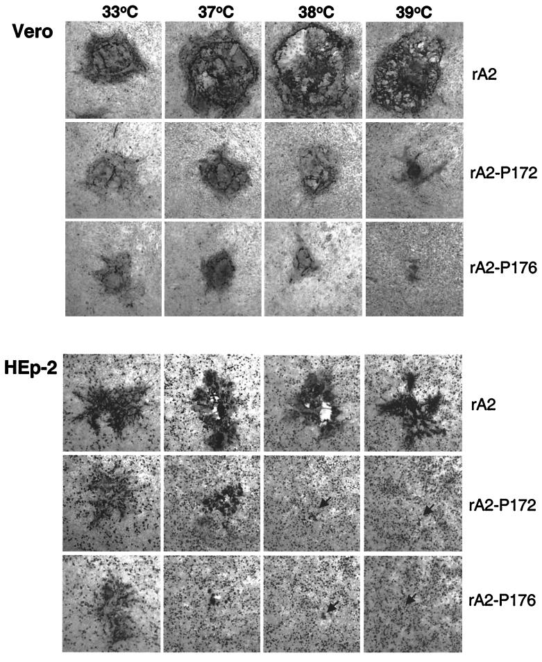
FIG. 4.
Plaque formation of rA2-P172 and rA2-P176 at different temperatures. Monolayers of Vero cells (upper panel) and HEp-2 cells (lower panel) were infected with wt rA2, rA2-P172, and rA2-P176; overlaid with L15 medium containing 1% methylcellulose and 2% FBS; and incubated at 33, 37, 38, and 39°C for 6 days. The plaques were visualized by immunostaining with polyclonal anti-RSV antibodies. Plaques were photographed on a Nikon inverted microscope. Arrows in the lower panels indicate RSV-infected HEp-2 cells at 38 and 39°C.

