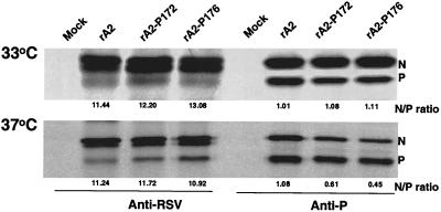
FIG. 6.
Immunoprecipitation of viral proteins from RSV-infected cells. Vero cells were infected with wt rA2, rA2-P172, or rA2-P176 at an MOI of 1.0 and incubated at 33 and 37°C for 18 h. Proteins were then radiolabeled with [35S]Cys and [35S]Met (100 μCi/ml) in DMEM deficient in cysteine and methionine for 4 h, immunoprecipitated by either anti-RSV or anti-P monoclonal antibodies, separated by SDS-15% PAGE, and autoradiographed. The positions of the N and P proteins are indicated on the right. The N and P ratio for each mutant was determined from four independent experiments.

