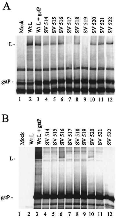
FIG. 1.
P-L complex formation with the Sendai virus L mutants. A549 cells in 35-mm-diameter dishes were infected with VVT7 at a multiplicity of infection of 2.5 PFU/ml and transfected with no plasmids (Mock) or Sendai virus gstP (0.2 μg) and the indicated wt or mutant Sendai virus L (1.67 μg) plasmids. The cells were incubated for 10 h and then labeled for 30 min using Express-35S (100 μCi/ml), and cytoplasmic extracts were prepared. Samples of the extracts were analyzed directly by sodium dodecyl sulfate-polyacrylamide gel electrophoresis (SDS-PAGE) for total protein expression (A) or incubated with glutathione-Sepharose beads, after which the bound proteins were separated by SDS-PAGE (B). The positions of the proteins are indicated.

