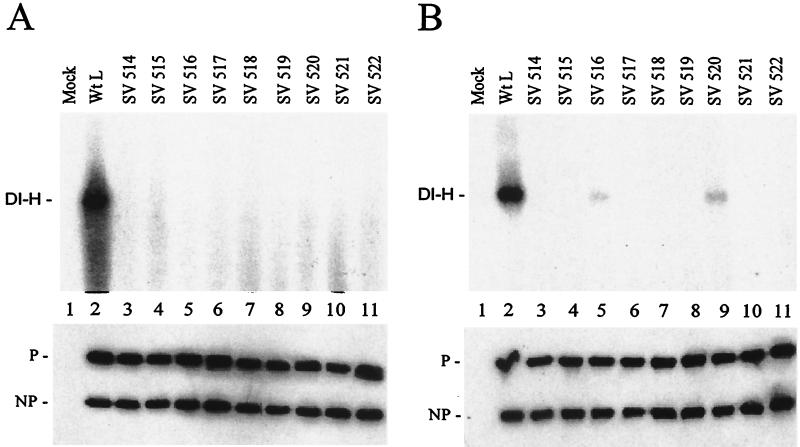
FIG. 5.
DI-H replication with the Sendai virus L mutants. (A) A549 cells were infected with VVT7 and transfected with no plasmids (Mock) or the Sendai virus NP, P, and the indicated wt or mutant L plasmids. Cytoplasmic extracts were incubated with polymerase-free DI-H RNA-NP in the presence of [α-32P]CTP, and total RNA was isolated and analyzed on an agarose-urea gel as described previously (8). The position of DI-H RNA is indicated. (B) For in vivo replication infected cells were transfected as above with the addition of pSPDI-H plasmid, and extracts were prepared. The extracts were nuclease treated, and the nuclease-resistant RNA was isolated and then separated on an agarose-urea gel. The RNA was transferred to a nylon membrane, and the blot was probed with a DI-H-specific (+) sense 32P-labeled riboprobe as described previously (8). The position of the DI-H RNA is indicated. (Bottom panels) Samples of the extracts were immunoblotted using α-SV and α-P antibodies. The positions of the proteins are indicated.

