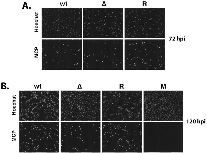
FIG. 12.
MCP expression in cultures infected with wt IE2 86-EGFP (wt), IE2 86ΔSX-EGFP (Δ), or IE2 86ΔSX-EGFP revertant (R) viruses. HFF cells were seeded on coverslips and infected with virus at an MOI of 0.5 in media containing 10% FBS. At the time points indicated, cells were fixed in 2% paraformaldehyde. After permeabilization, cells were stained with MCP-specific MAb followed by antimouse IgG TRITC-conjugated secondary antibody. Hoechst stain was used to visualize the total number of nuclei in the field. A field of mock-infected cells (M) was shown for comparison. Magnification, ×71.

