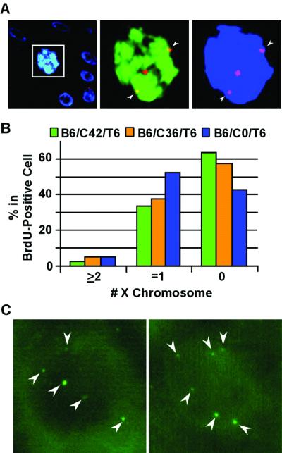
FIG. 3.
HPV-18 E7 induces endoreduplication in differentiated keratinocytes. Fluorescent images were taken from the differentiated strata in E7-transduced cultures. The labeling scheme is similar to that depicted in Fig 1A (top rows), except that the order of exposure to TdR analogues was reversed. FISH for panels A and B was conducted with cy3-conjugated, pericentromeric probes specific for the X chromosome (red), while BrdU was revealed with an FITC-conjugated anti-BrdU antibody (green) and nuclei were revealed with DAPI (in blue) in a 5-μm section. (A) B6/C42/T6 culture images were captured with a triple-pass filter (left) or with dual filters for the X chromosome and BrdU (middle) or for the X chromosome and the nucleus (right). Also note that the boxed nucleus is significantly larger than the nuclei of surrounding cells. (B) Percentages of differentiated cells containing 0, 1, or 2 or more X chromosome copies in BrdU-positive cells were scored in three E7-transduced cultures, B6/C42/T6, B6/C36/T6, and B6/C0/T6. (C) A FISH assay was conducted with fluorescein-conjugated pericentromeric probes of chromosome 17. Examples of six dots in a BrdU-negative nucleus from the B6/C36/T6 culture are shown. Weaker signal dots were not in focus because they were situated at different focal planes than the strong-signal dots. Arrowheads point to the pericentromeric signals.

