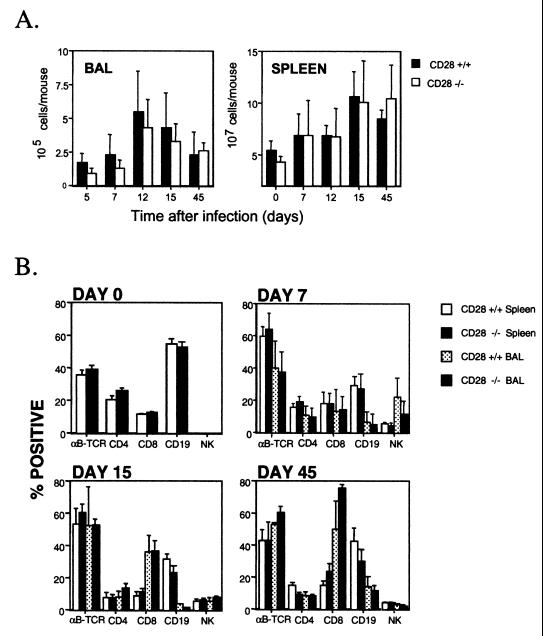
FIG. 2.
Cell numbers and lymphocyte subsets in the BAL or spleen of CD28+/+ and CD28−/− mice. (A) Cell numbers in the BAL or spleen were determined at intervals after intranasal infection of CD28+/+ and CD28−/− mice with MHV-68. Single-cell suspensions were prepared from the spleens of individual mice, as described previously (1), and viable cell counts were determined by trypan blue exclusion. Data are means + standard deviations of cell counts for three to eight mice at each time point. (B) BAL or spleen cells were stained with phycoerythrin- or fluorescein isothiocyanate-conjugated monoclonal antibodies, as previously described (25). The resulting populations were analyzed by flow cytometry. The detection limit was less than 1% based on staining with isotype-matched control antibodies. The means + standard deviations of data from two separate experiments at each time point are shown, with the exception of day 0, which was a single experiment. Groups of three to five mice were used at each time point. TCR, T-cell receptor.

