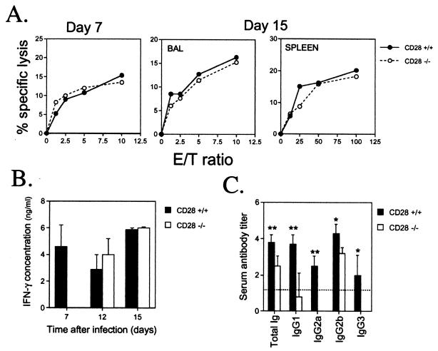
FIG. 3.
Cell-mediated and humoral immune responses to MHV-68 in CD28+/+ and CD28−/− mice. (A) Cytotoxic T-cell responses. Single-cell suspensions were prepared from BAL or spleens of individual mice at days 7 and 15 after intranasal infection with MHV-68 CTL activity was determined in a 6-h 51Cr release assay using MHV-68-infected MEF-1 cells as targets, as described previously (30). Mean percent specific lysis is shown for three separate experiments at day 7 and two at day 15. Groups of two or three mice were used in each experiment. No CTL activity was detected in the spleens of either CD28+/+ or CD28−/− mice at day 7 after infection. (B) Splenic recall IFN-γ responses. Spleens were harvested 15 or 30 days after infection with MHV-68. Splenocytes were restimulated with MHV-68-infected antigen-presenting cells, and supernatants were collected 24 to 96 h later for analysis of IFN-γ levels by sandwich ELISA, as previously described (25). Peak values (which usually occurred after 72 h in culture) are reported. Data are expressed as mean IFN-γ concentrations (nanograms/milliliter) + standard deviations for two separate experiments at day 7 and one each at days 12 and 15. Splenocyte cultures from three individual CD28+/+ or CD28−/− mice were tested in each experiment. (C) MHV-68-specific antibody responses. Serum was collected from CD28+/+ and CD28−/− mice 45 to 50 days after infection with MHV-68. Virus-specific antibody responses were determined by ELISA, as described previously (27). The titer of a serum sample was taken as the log10 of the highest dilution that gave an absorbance reading of <0.1. Data are expressed as mean serum antibody titers + standard deviations for two separate experiments. Sera from three individual mice were tested in each experiment. Asterisks denote that there was a statistically significant difference in antibody titers in CD28−/− and CD28+/+ mice. ∗, P < 0.05; ∗∗, P < 0.01 (Mann-Whitney rank sum test). E/T, effector-to-target.

