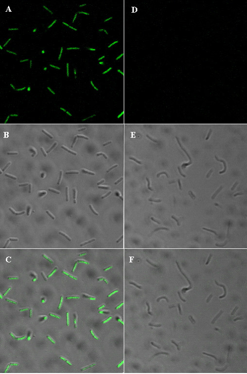
Figure 4.

Confocal scanning laser micrographs of GFP fluorescence of transformed Lactobacillus delbrueckii. (A – C) Genetically transformed lactobacillus with plasmid pLBS-GFP-EmR, and (D – F) control cells. Bacteria were grown overnight, washed, killed with sodium azide, and photographed under a laser scanning microscope (LSM) at scale of 54,8 × 54,8 μm. (C) Represents merged images of (A,B), and (F) represents merged images of (D,E).
