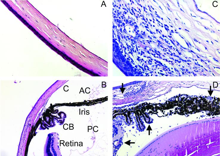
FIG. 5.
Histological analysis of the eye after HSV (KOS) infection. Normal uninfected tissue from C57BL/6 mice (A and B) and tissue from infected (129/SVEV × C57BL/6)F2 mice (C and D) were fixed in buffered 10% formalin, embedded in paraffin, and stained with hematoxylin and eosin. Infected mice showed corneal (C) infiltrates and swelling, neovascularization (arrows), and cellular infiltrates in the ciliary body (CB), iris, and anterior (AC) and posterior (PC) chambers. Magnification, ×40 (A, C, and D) or ×20 (B).

