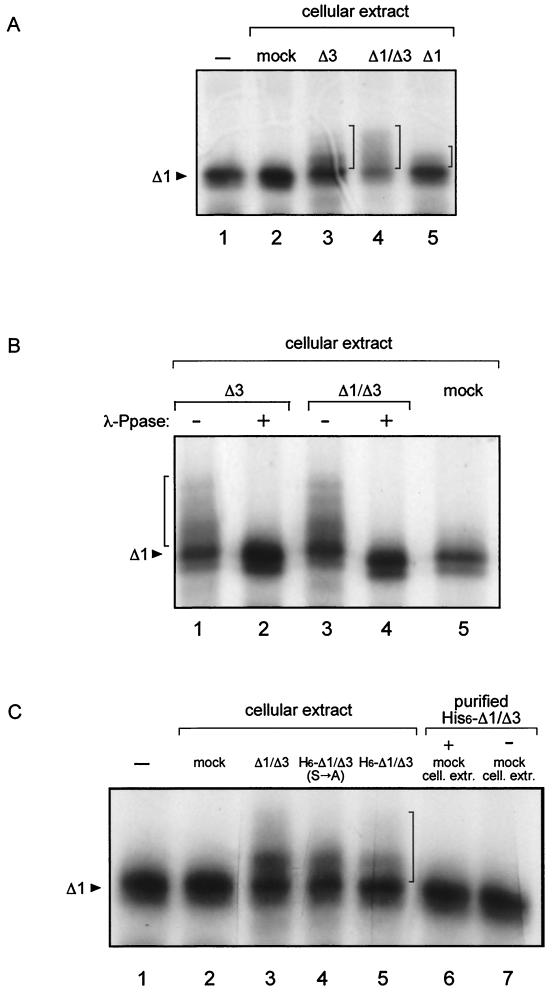
FIG. 2.
In vitro phosphorylation assay. Analysis of immunoprecipitates of in vitro-translated, [35S]methionine-labeled mutant Δ1. (A) Substrate was incubated with cellular extracts from cells transfected with the indicated mutants; mock, extracts from cells transfected with the same plasmid without the insert. −, no addition. (B) λ-Ppase treatment as indicated (+, treated; −, untreated). (C) Purified His6-Δ1/Δ3 was obtained by nickel column purification, and the same amount was used in lanes 5, 6, and 7. cell. extr., cellular extracts. The arrowheads indicate the unphosphorylated Δ1 substrate, and the vertical brackets indicate the positions of mobility-shifted phosphorylated forms. −, no addition.

