
Fig. 4.
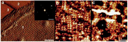
Organization of opsin in native Rpe65−/− disk membranes. a, three different surface types are discerned in the deflection image of a single-layered Rpe65−/− disk membrane: the paracrystalline, cytoplasmic surface of opsin (type 1), lipid (type 2), and mica (type 3). a, inset, calculated power spectrum of the paracrystalline region displayed in a. The first-order diffraction spot at (3.8 nm)−1 is marked by an arrow. b, the paracrystalline arrangement of opsin dimers (broken ellipse) in the native membrane. Occasional single opsin monomers are marked by arrowheads. c, the corrugated and flexible extracellular surface of opsin. The height between the lipid bilayer surface (asterisk) and clusters of opsin (triangle) is 2.8 ± 0.2 nm (n = 60). Scale bars: 50 nm (a), 5 nm−1 (a, inset), 15 nm (b), and 50 nm (c). Vertical brightness ranges: 0.3 nm (a), 1.6 nm (b), and 3.3 nm (c).