
Figure 4.
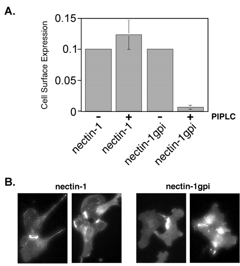
Cell surface expression and PIPLC sensitivity of GPI-linked nectin-1. (A) CELISA analysis. CHO-K1 cells expressing nectin-1 or nectin-1gpi were treated with PIPLC or mock treated, incubated with mAb CK6, followed by an antibody detection system. The assays were performed in triplicate and repeated two times with similar results. The mean values plus standard deviations for a representative experiment are depicted. The absence of error bars for mean values given is due to standard deviations too small to generate visible error bars. (B) Indirect immunofluorescence of cells expressing nectin-1α or nectin-1gpi. B78H1 cells expressing nectin-1α, B78H1 CJ4E, or nectin-1gpi, B78H1 n1gpiA, were fixed with ice-cold methanol and stained with anti-nectin-1 mAb CK41 followed by an FITC-conjugated secondary antibody.