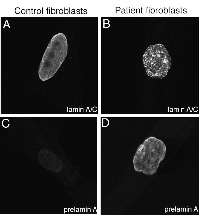
Figure 2. Immunofluorescent localization of lamins A and C and prelamin A in control and RD patient fibroblasts.

(A) Confocal micrographs show localization of lamins A and C in nuclei of a control fibroblast and (B) a fibroblast from a RD patient. (C) Confocal micrographs show staining for prelamin A in a control fibroblast and (D) in a fibroblast from a RD patient.
