
Figure 6.
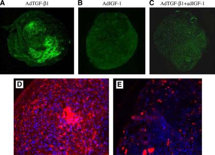
Detection of collagen type II and type X collagen expression in the pellets by immunofluorescence. (A): Pellet with AdTGF-β1-transduced cells at 50 MOI; (B): with AdIGF-1 at 150 MOI; (C): with combination of AdTGF-β1 and AdIGF-1 at 150 MOI (×100 magnification). Type II collagen immunostaining is present in the AdTGF-β1 pellets only. (D): Immunofluorescence for type X collagen in pellets made from nontransduced cells and incubated in a medium supplemented with TGF-β1 protein at 50 ng/mL. (E): Immunofluorescence for type X collagen in pellets made from MSCs transduced with AdTGF-β1 at 50 MOI. There is extensive staining for type X collagen in pellet D than in pellet E. The figure is representative of 3 stained pellets.