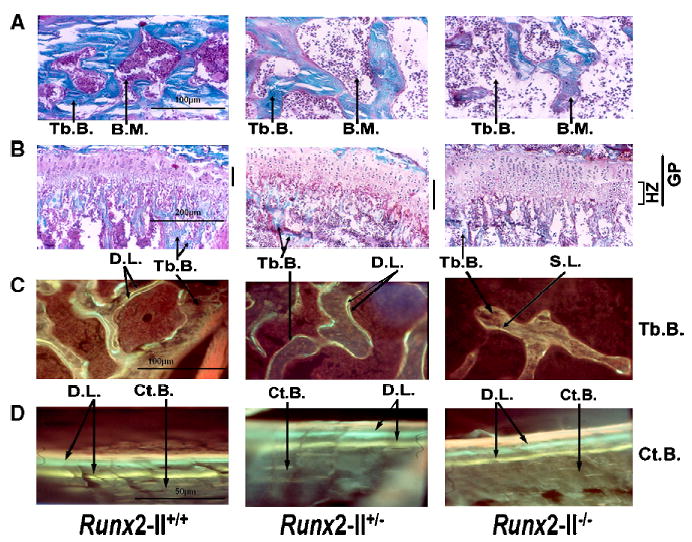
Fig. 4.

Nondecalcified histological sections of tibia bone in 6-week-old Runx2-II wild-type and mutant mice. (A, B) Goldner-stained sections of the epiphyseal bone (×200 magnification) and growth plate and metaphyseal bone (×100 magnification) of 6-week-old Runx2-II+/+, Runx2-II+/−, and Runx2-II−/− mice. Mineralized trabecular bone (Tb) is blue and unmineralized osteoid is reddish brown in color. (C, D) Villanueva-stained sections of trabecular (×200 magnification) and cortical bone (×400 magnification) viewed under fluorescent light in 6-week-old Runx2-II+/+, Runx2-II+/−, and Runx2-II−/− mice prelabeled with tetracycline followed by calcein (double label). Heterozygous Runx2-II+/− mice have reduced trabecular bone volume, normal appearing growth plate, and reduced bone formation rates. Homozygous Runx2-II−/− mice show a marked reduction of trabecular bone volume, a widened growth plate consisting of increased zone of hypertrophic chondrocytes, and severe impairment of bone formation as evidenced by a diffuse single label or unlabeled trabecular bone surfaces. In contrast, mineral apposition rates on the periosteal surfaces of cortical bone were preserved in both heterozygous Runx2-II+/− and homozygous Runx2-II−/− mice. Tb.B., trabecular bone, Ct.B., cortical bone, B.M., bone marrow, G.P., growth plate, H.Z., hypertrophic zone, D.L., double label, S.L., single label.
