
Fig. 3.
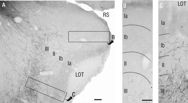
Details of PHA-L-labeled axons in rostral APC. A: Low-power photomicrograph showing PHA-L-labeled axons predominantly within layer III in the ventral half of rostral APC. B,C: High-power photomicrographs of the areas outlined in A. B: In dorsal APC, note the relatively small number of labeled fibers in layer III, and the absence of fibers in layers I and II. This contrasts remarkably with ventral APC (C), where PHA-L-labeled fibers were found in layers Ib, II, and III. Scale bars = 100 μm in A; 50 μm in B (applies to B,C).