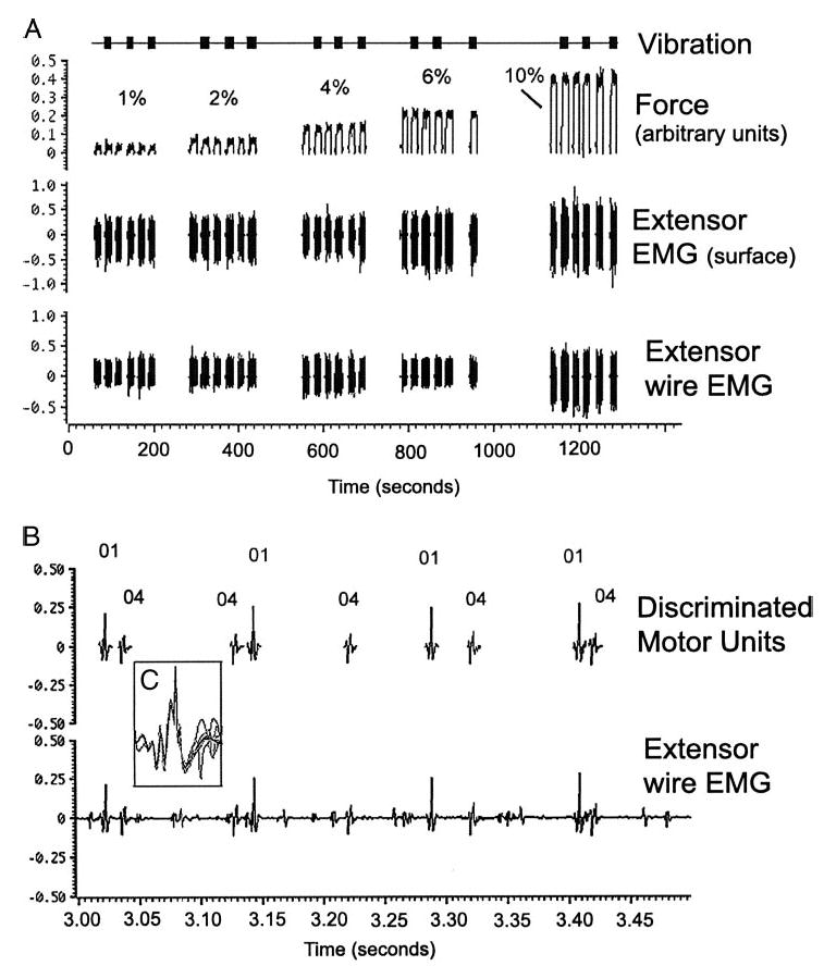
Fig. 1.

Example of the experimental procedure (patient 4). A: wrist extension is performed at a steady level for 10-s epochs with several seconds rest between contractions. In this example, there are 6 10-s epochs at each of 5 force levels: 1, 2, 4, 6, and 10% of maximal voluntary contraction. Top: timing of the muscle vibration. Middle and bottom: extensor muscle surface electromyography (EMG) and recordings from intramuscular wires. B: expanded scale (sweep = 500 ms) shows motor unit recordings and spike discrimination of 2 units during a contraction epoch at 10% maximal force. Both units were initially recruited at 1% force with firing frequencies of 9.7 and 11.3 Hz, similar to that seen at 10% force. Note that not all motor units visible in the recording were discriminated. C: inset showing overlay of 10 spikes discriminated by their morphology.
