
Fig. 8. TGF-β1-induced protection from serum deprivation-induced apoptosis of lung fibroblasts is dependent on the PI3K/Akt pathway.
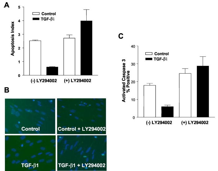
A, IMR-90 cells were grown to near-confluence in a 96-well ELISA culture plate and growth-arrested for 48 h in 0.01% serum prior to treatment with TGF-β1 ± LY294002 (10 μ m) as indicated for 5 days. Apoptosis rates were measured using an ELISA for single-stranded DNA as described under “Materials and Methods.” Findings represent one of three independent experiments. Values are mean ± S.E., n = 3. *, p < 0.05 compared with untreated controls. B, cultured IMR-90 cells were grown to 50% confluence, growth-arrested in 0.01% serum for 48 h, and then treated with/without TGF-β1 (2 ng/ml) in the presence/absence of the PI3K inhibitor, LY294002 (10 μ m), as indicated. Immunofluorescence staining for activated caspase 3 was performed 5 days after treatment. Counter staining with DAPI (blue) was used to identify nuclear morphology. C, quantitative assessment of apoptosis by activated caspase 3-positivity for the experimental conditions shown in B. Three random 20× fields containing 50–60 cells per field were selected from each group. % apoptosis was measured by determining dividing the total number of activated caspase 3-positive cells (fluorescein isothiocyanate/green) by the total number of cells (DAPI/blue). Findings are representative of three independent experiments. Values are mean ± S.E., n = 3. *, p < 0.05 compared with untreated controls.