
FIGURE 11.
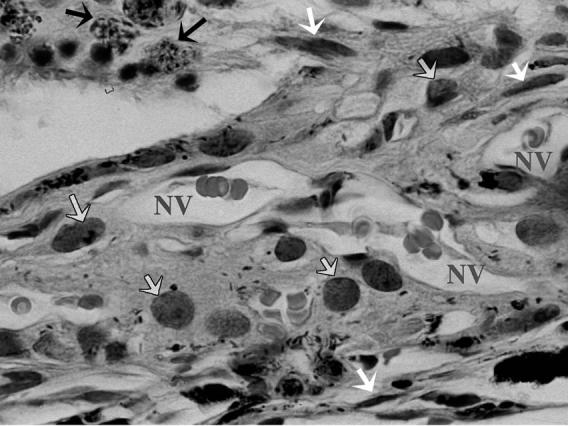
High Magnification Showing the Structure of Induced Choroidal Neovascular Membrane in One of the Control Eyes 4 Weeks After Laser Consisting of Proliferating Endothelial Cells Forming New Vessels (NV) Containing RBCs, Spindle-Shaped Cells (fibroblasts) Pointed to by White Arrows, Macrophages (Gray Arrows), and Pigment Laden Cells (Black Arrows). (H & E stain, original magnification X = 330).