
FIGURE 3.
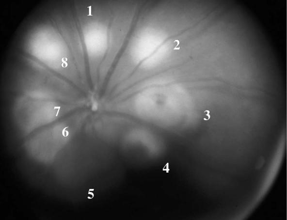
Fundus Photo of One of the Treated Eyes of the Pretreatment Group Captured 1 Day After Laser. Lesions 1, 2, and 8 were associated with bubbling. Lesions 6 and 7 were associated with choroidal hemorrhage. Lesions 3 and 4 were associated with retinal hemorrhage. Spot 5 was associated with preretinal hemorrhage.