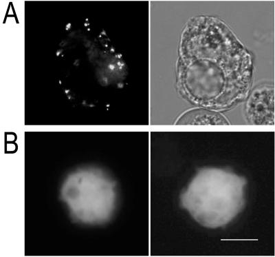
FIG. 2.
(A) Fluorescence (left) and light (right) micrographs of a live Acanthamoeba expressing the full-length AhSub-EGFP fusion protein. Small fluorescent vesicle-like structures were distributed at the periphery of the cytoplasm. (B) Two amoebae with the fluorescence of EGFP alone showed dispersed distribution in the cytoplasm. Bar, 10 μm.

