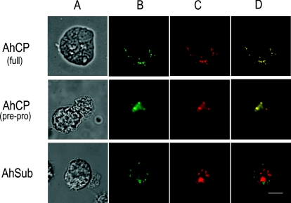
FIG. 6.
Localization of AhCP-EGFP in A. healyi. (A) Light micrograph; (B) expressed AhCP-EGFP and AhSub-EGFP fusion proteins (green signal); (C) lysosomes of Acanthamoeba stained with Lyso Tracker Red DND-99 (red signal); (D) overlapped micrograph images of panels B and C (yellow signal). The presence of the yellow signals was demonstrated only in the cells expressing AhCP but not in the cells transfected with AhSub. Bar, 10 μm.

