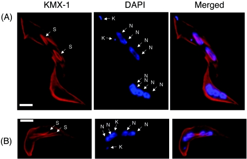
FIG. 6.
Mitotic spindle labeling in TbPLK-depleted cells. Cells 2 days into RNAi were fixed with 3.7% formaldehyde and labeled with KMX-1 and DAPI. (A) Two tetranucleated cells. The one above has two short mitotic spindle structures (S) each mediating a pair of nuclei (N), suggesting the state of metaphase. The one below shows no spindle structure. (B) Tetranucleated cell with two elongated spindle structures each connected to a pair of nuclei. It suggests that the cell may be in late anaphase. K, kinetoplast. Bars, 5 μm.

